Pelvic Female Abdomen Ultrasound - Pelvic Ultrasound Johns Hopkins Medicine / This ultrasound examination will examine all the abdominal organs visible on ultrasound scanning.
byAdmin•
0
Pelvic Female Abdomen Ultrasound - Pelvic Ultrasound Johns Hopkins Medicine / This ultrasound examination will examine all the abdominal organs visible on ultrasound scanning.. How to prepare for the test. There are three types of pelvic ultrasound: It allows your doctor to see your bladder, cervix, uterus, fallopian tubes, and ovaries. A female pelvis ultrasound, a.k.a. Your doctor is easily able to view the uterus, cervix, vagina, fallopian tubes and ovaries during a pelvic ultrasound.
Depending on the reason for the test, women also may have a transvaginal ultrasound during the same visit. Ultrasound imaging uses soundwaves to create pictures of the inside of the body. Pelvic ultrasound can help to diagnose a variety of conditions in both men, women, and children. The test is painless and easy to tolerate. An abdominal ultrasound is used to evaluate the organs and blood vessels in the abdomen, including the gallbladder, kidneys, liver, pancreas and spleen.
Abdominal Pain In Nonpregnant Female Patients 2014 04 06 Ahc Media Continuing Medical Education Publishing from www.reliasmedia.com For instance, a pelvic ultrasound allows a doctor to take images of the female organs such as the ovaries cervix, uterus, and fallopian tubes, as well as the images of male organs, including the prostate gland and seminal vesicles. In women, pelvic pain may be a sign of menstrual cramps, ovulation, or a gastrointestinal issue such as a. An ultrasound of the pelvis is typically used to look at the bladder, ovaries, uterus, cervix, and fallopian tubes (some of these are known as the female reproductive organs). An ultrasound of the abdomen or pelvis can identify gallstones, an infected gallbladder, thickening of the gallbladder wall, and excess fluid surround the gallbladder. Ultrasound imaging uses soundwaves to create pictures of the inside of the body. Please follow these simple instructions so that we may better serve you. Transabdominally (through the abdomen) and transvaginally (through the vaginal canal). Your doctor might order this test to diagnose a condition, or to check the health of your baby.
In women, pelvic pain may be a sign of menstrual cramps, ovulation, or a gastrointestinal issue such as a.
However, its views may be limited by abdominal structures such as bowel gas. The sound waves create a picture on a video monitor. Pelvic ultrasound scans are regularly deployed to identify any causes of these symptoms such as uterine polyps, fibroids, ovarian cysts. This is a condition where the endometrial tissue located inside the uterus cavity, migrates to other parts of the pelvis, such as the tubes, ovaries, abdominal. Generalised abdominal and pelvic pain, discomfort, bloating, with or without abnormal vaginal bleeding, lower back pain, kidney region, blood in the urine, and urine infection are the most common reasons for having an abdominal, pelvic and urinary tract. A pelvic ultrasound is a test that uses sound waves to make pictures of the organs inside your pelvis. You may be asked to drink a few glasses of water to fill your bladder. A renal ultrasound is used to evaluate the kidneys, ureters and bladder. Your doctor has requested that an ultrasound exam be performed on your abdomen and pelvis. Pelvic pain (most comon) swelling or a bloated feeling of the abdomen. Abdominal, or transabdominal ultrasounds can produce images of the bladder, uterus, cervix, ovaries and fallopian tubes. The test is painless and easy to tolerate. Liver, gallbladder, kidneys, bladder, uterus, ovaries, prostate and seminal vesicles.
In women, pelvic pain may be a sign of menstrual cramps, ovulation, or a gastrointestinal issue such as a. Please follow these simple instructions so that we may better serve you. A pelvic ultrasound may be done with a full bladder. The test is painless and easy to tolerate. If your doctor orders a pelvic ultrasound exam, images can be captured in two different ways:
Gynecology Pelvic Ultrasound Made Easy Step By Step Guide Pocus 101 from pocus101.b-cdn.net A female pelvis ultrasound, a.k.a. The abdominal, the pelvic/gynaecology and the urinary ultrasound scan. An ultrasound of the pelvis is typically used to look at the bladder, ovaries, uterus, cervix, and fallopian tubes (some of these are known as the female reproductive organs). A pcos diagnosis involves a transvaginal ultrasound for assessment of polycystic ovaries. In women, pelvic pain may be a sign of menstrual cramps, ovulation, or a gastrointestinal issue such as a. Excretory organs like the kidneys, urethra, bladder and bowel, and surrounding structures like blood vessels. Imaging tests can identify abnormalities and help doctors diagnose conditions. Some symptoms of pelvic mass may include:
This ultrasound examination will examine all the abdominal organs visible on ultrasound scanning.
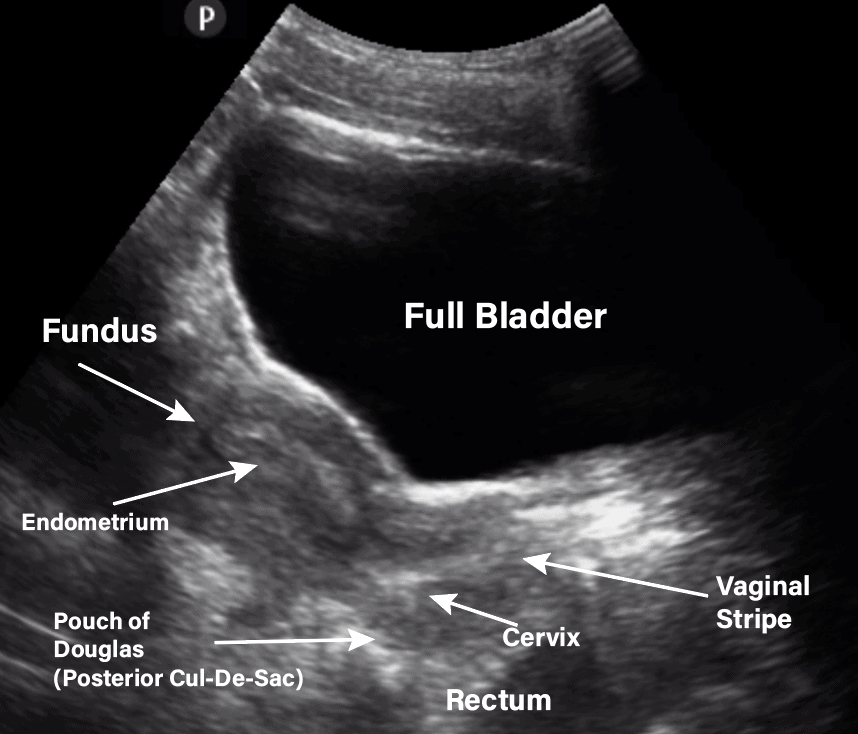
An ultrasound, also named sonography, of the abdomen and the pelvic makes it possible to see your abdominal and pelvic organs: The role of pelvic ultrasound. Generalised abdominal and pelvic pain, discomfort, bloating, with or without abnormal vaginal bleeding, lower back pain, kidney region, blood in the urine, and urine infection are the most common reasons for having an abdominal, pelvic and urinary tract. A pelvic ultrasound may be done with a full bladder. Please follow these simple instructions so that we may better serve you. Excretory organs like the kidneys, urethra, bladder and bowel, and surrounding structures like blood vessels. For instance, a pelvic ultrasound allows a doctor to take images of the female organs such as the ovaries cervix, uterus, and fallopian tubes, as well as the images of male organs, including the prostate gland and seminal vesicles. A pelvic ultrasound is a test that uses sound waves to make pictures of the organs inside your pelvis. 1.) the first part is to look at the pelvic organs from the outside of your abdomen called transabdominal pelvic ultrasound. However, its views may be limited by abdominal structures such as bowel gas. Having a full bladder can help with looking at organs, such as the womb (uterus), within your pelvis. Your doctor has requested that an ultrasound exam be performed on your abdomen and pelvis. Pelvic ultrasound can help to diagnose a variety of conditions in both men, women, and children.
Depending on the reason for the test, women also may have a transvaginal ultrasound during the same visit. Excretory organs like the kidneys, urethra, bladder and bowel, and surrounding structures like blood vessels. An abdominal ultrasound is used to evaluate the organs and blood vessels in the abdomen, including the gallbladder, kidneys, liver, pancreas and spleen. Your doctor might order this test to diagnose a condition, or to check the health of your baby. Abdominal, or transabdominal ultrasounds can produce images of the bladder, uterus, cervix, ovaries and fallopian tubes.
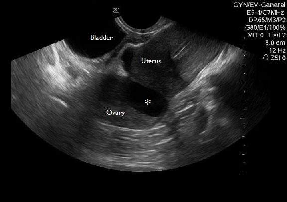
18 Ultrasound Ideas Ultrasound Sonography Youtube from i.pinimg.com Many women may have a pelvic mass at some point in their live, although not all women will experience sympotms. These exams are frequently used to evaluate the reproductive and urinary systems. A pelvic ultrasound may be done with a full bladder. Excretory organs like the kidneys, urethra, bladder and bowel, and surrounding structures like blood vessels. It's the preferred screening method for an abdominal aortic aneurysm, a weakened, bulging spot in the abdominal aorta — the major blood vessel that supplies blood to the body. A pelvic ultrasound allows quick visualization of the female pelvic organs and structures including the uterus, cervix, vagina, fallopian tubes and ovaries. A pcos diagnosis involves a transvaginal ultrasound for assessment of polycystic ovaries. There are three types of pelvic ultrasound:
A pelvic ultrasound may be done with a full bladder.
How to prepare for the test. Transabdominally (through the abdomen) and transvaginally (through the vaginal canal). Abdominal, or transabdominal ultrasounds can produce images of the bladder, uterus, cervix, ovaries and fallopian tubes. The role of pelvic ultrasound. A pelvic ultrasound is a noninvasive diagnostic exam that produces images that are used to assess organs and structures within the female pelvis. The sound waves create a picture on a video monitor. Pelvic ultrasound imaging has two components. London private ultrasound (lpu) offers a combined abdominal, pelvic, and urinary tract ultrasound scan to female patients over 18 years old. An abdominal ultrasound is used to evaluate the organs and blood vessels in the abdomen, including the gallbladder, kidneys, liver, pancreas and spleen. In general for pocus exams, it is usually good to start with the transabdominal ultrasound and then use the transvaginal approach if needed. Depending on the reason for the test, women also may have a transvaginal ultrasound during the same visit. Abdominal, vaginal (for women), and rectal (for men). 1.) the first part is to look at the pelvic organs from the outside of your abdomen called transabdominal pelvic ultrasound.
Pelvic pain (most comon) swelling or a bloated feeling of the abdomen pelvic ultrasound female. However, its views may be limited by abdominal structures such as bowel gas.